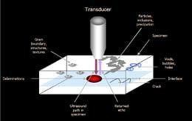
The scientific name is: Scanning Acoustic Microscope, which works on the principle of reflection and transmission of sound wave pulses. Its core is a microwave chain with piezoelectric ceramics. Piezoelectric ceramics generate short sound pulses, which are then focused together by the acoustic lens. The main component of the scanning acoustic microscope is called the transducer, which is also the device that generates ultrasonic signals together with the acoustic lens.
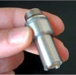
Normal transducer
Another mode for ultrasonic analysis is called transmission mode. During transmission scanning, another transducer is installed under the sample, which will receive all ultrasonic signals that completely penetrate the sample. Ultrasonic images can be generated based on the received signals.
KSI high-speed low-noise patented transducer
In order to generate a complete sample image, a line-by-line scanning method is adopted. The transducer acoustic axis on the transducer has good focusing performance and can both send and receive signals. The sample is scanned in this line-by-line and point-by-point manner to form an ultrasonic image. The scanning time to generate a complete image depends on the size of the scanned sample and the resolution of the image. Generally speaking, an ultrasonic image of a small sample can be completed in just a few seconds.Working principle
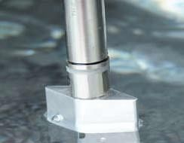
The transducer is responsible for converting electromagnetic pulses into acoustic pulses. After leaving the transducer, the sound field is focused on the sample by the acoustic lens through the medium (deionized water or anhydrous alcohol, etc.). Water is a coupling medium to prevent the attenuation of ultrasonic signals. The sample is placed in water. As long as the sound wave signal encounters acoustic impedance (such as pores, bubbles, etc.) on the surface or inside of the sample, reflection will occur. After the transducer receives the reflected signal, it will convert it into an electrical pulse. After the ultrasonic signal is converted into an electrical pulse, it is represented by 256 grayscale values. Each transducer has its own specific ultrasonic frequency, which can be specially configured according to the needs of the user. This process is the basic process of the reflection working mode of the scanning acoustic microscope.